
What is the wellness mini trampoline lymphacising
health-bounce?
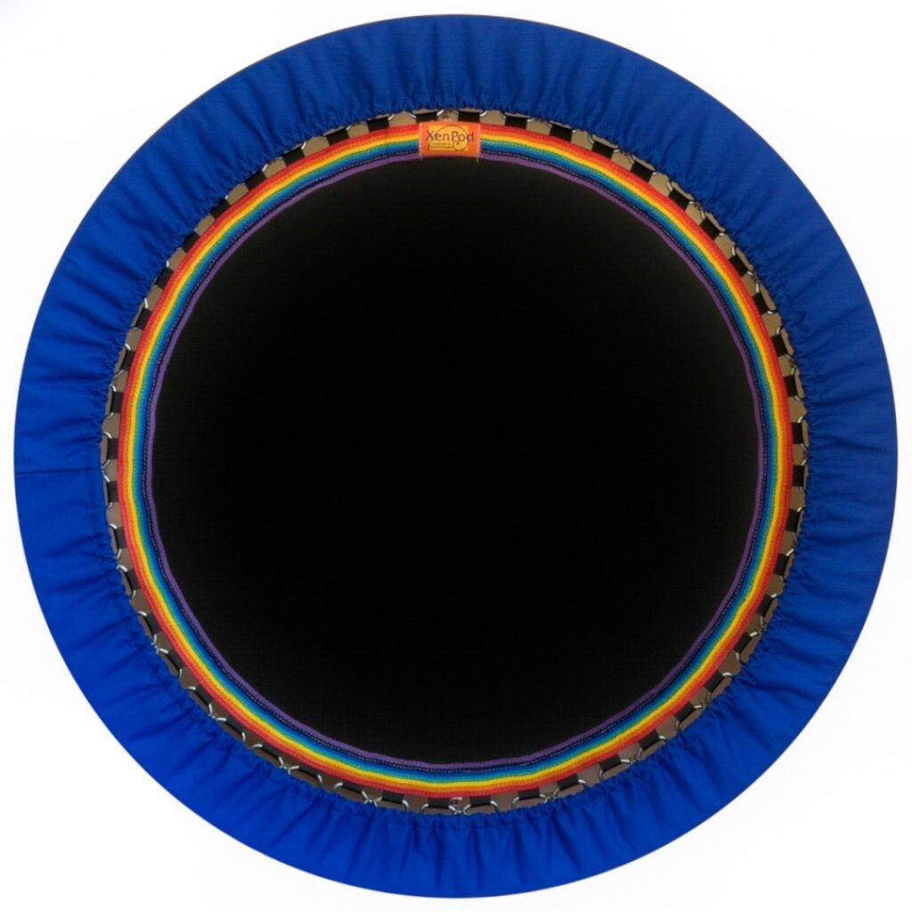
The pioneers of lymphacising discovered that there is a very effective way to assist the lymphatic system to function optimally, by using a very specific type of bounce: the healing health-bounce This is why XenPod’s innovator Ian Pettitt spent his life working to precisely calibrate the XenPod to give what we call the perfect health-bounce.
This exact calibration creates the correct G-forces needed to efficiently shunt lymph fluid upwards through one way valves in the lymph system at an increased rate. This accelerated movement of lymph will then find it’s way to the lymphatic drainage ducts in the torso and out of the body via the five elimination systems – skin, lungs, kidneys, liver and bowel.
The healing health-bounce uses the gravitational G force of acceleration and deceleration to create heat and pressure that charges the polarity and chemical make-up of weak cells. This activates the cells sodium potassium pump (ATP) which is responsible for balancing the correct electrical function of individual cells and fluids, to maintain positive blood pressure and negative lymph sub-pressure.

For anyone, any time, anywhere
With regular lymphacising every person young or old can aim for the optimum cellular ‘dry state’ where all the wastes have been removed. As little as 20 minutes per day while watching a movie or seminar, or listening to your favourite music will give great benefits.


Self-Heal with Lymphacising

The Lymphatic System is the foundation of the immune, digestive, cleansing and cell communication systems. It controls the body’s energy and information circuits, enzyme production and regeneration.
If the lymphatic system becomes sluggish or impaired in its task of removing cellular waste, the information it carries will be overloaded with toxins and the body will become unhealthy. Therefore if we can assist the body in moving lymph fluid more efficiently with a lymphaciser, our health will improve and pain will subside.
The lymph vessel network has thousands of one-way valves that pump lymph fluid upwards against gravity as the body moves. Every day as we walk and go about our daily work, the body will attempt to keep the lymph moving. Unfortunately due to toxic overload and the force of gravity, our normal movements each day may often be insufficient to completely remove all wastes and cells will be constantly in what is known as the ‘wet state’ – where there is excess fluid present that traps blood proteins in a toxic acidic environment. These toxic conditions facilitate disease, allow infections and create complications like lymphoedema. They adversely affect the lymphatic system, which if not properly cleansed will become more and more congested, sluggish, overwhelmed and diseased.
The benefits of Lymphacising
Gain a healthier body, mind and spirit and have more energy for life! Create free access to your own holistic health-care while at home or at work. Self-correct your own posture daily to increase flexibility, functionality and wellbeing.
Compliment all your sports, therapies and body building gym routines and work at your own pace
You can powerfully help yourself, to reverse body injury and pain
Enter deep relaxation and meditation when lying on the XenPod in the peaceful harmonic resonance
Stimulate a better immune defence by maintaining a healthier lymphatic system
Ideal for the busy person. Multi-task by using your XenPod when you’re doing other things
Do away with expensive, recurring treatments with this quality daily self care machine
Increase oxygen at cell level, by generating more A.T.P. with the healing health bounce
Less sleep is required for a days work when lymphacising regularly
Dr Ez video about health, lymphology and what happens when you health-bounce
Dr West (Jr) DL PMD DD is a firm advocate of the healing health-bounce and he gives many lectures on the science of Lymphology and how critical this knowledge is: in creating health or disease. He covers the cellular ‘Dry State’ of good health and immunity, the cell fuel of ATP and the vital Sodium Potassium power pump, which his father Dr Samuel C. West discovered. Here he explains how bouncing is key in maintaining this healthy cellular ‘Dry State’.
⚠️ Beware of using cheap rebounders!
Bouncing on low-quality rebounders can harm your health, especially as you age. Test this with kinesiology: check your muscle strength before and after using the rebounder. If you're weaker after bouncing, the machine may be impacting your health negatively. XenPod's advanced equipment ensures a safe and revitalising experience for your wellness journey.
Learn about your lymphatic and body fluid systems
The Four Vital Circulatory Systems
The action of lymphacising powerfully affects the four vital fluid systems of the body to cleanse, balance and vitalise all fluids for optimal health.
The four vital circulatory systems consist of the oxygenated red blood flow, the dark waste venous flow, the cerebral spinal fluid flow and the lymphatic fluid flow. Of these four, the lymphatic system has by far the greatest volume of fluid to move around the body and requires the greatest encouragement to move.

1. Blood Flow
The first system is well documented as the circulatory or the red blood system. After getting oxygen and hydrogen from the lungs, oxygenated blood containing iron and other organic elements, is pumped by the heart via its vascular systems: basically taking our life-force to all the cells of our body.

2. Venous Flow
The second system is the venous system which carries the de-oxygenated (dark) blood back to the heart to process cellular wastes and gases. The lungs take away gaseous wastes allowing the next breath to receive another charge of oxygen, hydrogen and elements from the heart: to once again supply the cells needs. If the waste produced by the cells were returned to the blood, or not processed enough, we would suffer from toxemia, which is poisoning of the blood. Therefore the body has to have a return system, much like the white blood system, which removes the cellular waste and gases from our body.
The venous system extends to every cell and drains away cellular wastes. It is returned to the blood system after purification via the kidneys and liver. Purified blood is then returned to the circulatory system to continue the never ending process.

3. Spinal Fluid
The third system is the cerebral spinal fluid system. This slow moving fluid travels through all the cranium plates, the spine and sacrum and extends to all joints in the body. It takes wastes and toxins from the central nervous system, down the spine to the sacral pump which pumps up nutrients needed for the central nervous and cranial muscle systems.
Effective cleansing is accelerated with the Hip Aligning and Step Breath done in the XenPod program. Each small breath is synchronised with the bottom of each bounce. Maximum chest expansion and spine flexion assures the sacrum will attain maximum range of motion for the cerebral spinal fluid to speed-up the central nervous system’s huge responsibility of regenerating the self-healing human organism.

4. Lymph ~ The White Blood Flow
The fourth and largest system is the lymphatic fluid, which is like a slow white blood flow. The incredible lymph system works in harmony with the red blood system, with every blood vessel having a corresponding lymph vessel. The lymph has five times more volume than blood in the body and moves much more slowly.
The cells of our body are nourished by oxygen, minerals and organic nutriments obtained from the food we eat every day. All systems of the body gain access to these vital nutrients via our lymph fluid and the elements, enzymes and vital communication processes that it facilitates. You can see this clear fluid leaking from a burn, or from a common blister. The science of Lymphology explains how lymphacising removes trapped blood protein wastes from the lymph fluid and this science is the basis of our Energising while Lymphacising© program.
The Lymphatic System FYI Extracted from Gray’s Anatomy
Henry Gray (1821–1865). Anatomy of the Human Body. 1918.
Introduction
THE LYMPHATIC SYSTEM consists (1) of complex capillary networks which collect the lymph in the various organs and tissues; (2) of an elaborate system of collecting vessels which conduct the lymph from the capillaries to the large veins of the neck at the junction of the internal jugular and subclavian veins, where the lymph is poured into the blood stream; and (3) lymph glands or nodes which are interspaced in the pathways of the collecting vessels filtering the lymph as it passes through them and contributing lymphocytes to it. The lymphatic capillaries and collecting vessels are lined throughout by a continuous layer of endothelial cells, forming thus a closed system. The lymphatic vessels of the small intestine receive the special designation of lacteals or chyliferous vessels; they differ in no respect from the lymphatic vessels generally excepting that during the process of digestion they contain a milk-white fluid, the chyle.
The Development of the Lymphatic Vessels.
The lymphatic system begins as a series of sacs at the points of junction of certain of the embryonic veins. These lymph-sacs are developed by the confluence of numerous venous capillaries, which at first lose their connections with the venous system, but subsequently, on the formation of the sacs, regain them. The lymphatic system is therefore developmentally an offshoot of the venous system, and the lining walls of its vessels are always endothelial.
In the human embryo the lymph sacs from which the lymphatic vessels are derived are six in number; two paired, the jugular and the posterior lymph-sacs; and two unpaired, the retroperitoneal and the cisterna chyli. In lower mammals an additional pair, subclavian, is present, but in the human embryo these are merely extensions of the jugular sacs.
The position of the sacs is as follows: (1) jugular sac, the first to appear, at the junction of the subclavian vein with the primitive jugular; (2) posterior sac, at the junction of the iliac vein with the cardinal; (3) retroperitoneal, in the root of the mesentery near the suprarenal glands; (4) cisterna chyli, opposite the third and fourth lumbar vertebrae (see fig. 592 above). From the lymph-sacs the lymphatic vessels bud out along fixed lines corresponding more or less closely to the course of the embryonic blood vessels. Both in the body-wall and in the wall of the intestine, the deeper plexuses are the first to be developed; by continued growth of these the vessels in the superficial layers are gradually formed. The thoracic duct is probably formed from anastomosing outgrowths from the jugular sac and cisterna chyli. At its connection with the cisterna chyli it is at first double, but the two vessels soon join. All the lymph-sacs except the cisterna chyli are, at a later stage, divided up by slender connective tissue bridges and transformed into groups of lymph glands. The lower portion of the cisterna chyli is similarly converted, but its upper portion remains as the adult cisterna.
Lymphatic Capillaries
The complex capillary plexuses which consist of a single layer of thin flat endothelial cells lie in the connective-tissue spaces in the various regions of the body to which they are distributed and are bathed by the intercellular tissue fluids. Two views are at present held as to the mode in which the lymph is formed: one being by the physical processes of filtration, diffusion, and osmosis, and the other, that in addition to these physical processes the endothelial cells have an active secretory function. The colorless liquid lymph has about the same composition as the blood plasma. It contains many lymphocytes and frequently red blood corpuscles. Granules and bacteria are also taken up by the lymph from the connective-tissue spaces, partly by the action of lymphocytes which pass into the lymph between the endothelial cells and partly by the direct passage of the granules through the endothelial cells.
The lymphatic capillary plexuses vary greatly in form; the anastomoses are usually numerous; blind ends or cul-de-sacs are especially common in the intestinal villi, the dermal papillæ and the filiform papillæ of the tongue. The plexuses are often in two layers: a superficial and a deep, the superficial being of smaller caliber than the deep. The caliber, however, varies greatly in a given plexus from a few micromillimeters to one millimeter. The capillaries are without valves.
Distribution - The Skin.
Lymphatic capillaries are abundant in the dermis where they form superficial and deep plexuses, the former sending blind ends into the dermal papillæ. The plexuses are especially rich over the palmar surface of the hands and fingers and over the plantar surface of the feet and toes. The epidermis is without capillaries. The conjunctiva has an especially rich plexus. The subcutaneous tissue is without capillaries.
The tendons of striated muscle and muscle sheaths are richly supplied. In muscle, however, their existence is still disputed. The periosteum of bone is richly supplied and they have been described in the Haversian canals. They are absent in cartilage and probably in bone marrow. The joint capsules are richly supplied with lymphatic capillaries, they do not, however, open into the joint cavities. Beneath the mesothelium lining of the pleural, peritoneal and pericardial cavities are rich plexuses; they do not open into these cavities. The alimentary canal is supplied with rich plexuses beneath the epithelium, often as a superficial plexus in the mucosa and a deeper submucosal plexus. Cul-de-sacs extend into the filiform papillæ of the tongue and the villi of the small intestine. Those portions of the alimentary canal covered by peritoneum, have in addition a subserous lymphatic capillary plexus beneath the mesothelium.
Other Organs and their part in the Lymphatic System
The salivary glands are supplied with lymphatic capillaries.
The liver has a rich subserous plexus in the capsule and also extensive plexuses which accompany the hepatic artery and portal vein. The lymphatic capillaries have not been followed into the liver lobules. The lymph from the liver forms a large part of that which flows through the thoracic duct. The gall-bladder and bile ducts have rich subepithelial plexuses and the former a subserous plexus.
The spleen has a rich subserous set and a capsular set of lymphatic capillaries. Their presence in the parenchyma is uncertain.
The nasal cavity has extensive capillary plexuses in the mucosa and submucosa.
The trachea and bronchi have plexuses in the mucosa and submucosa but the smaller bronchi have only a single layer. The capillaries do not extend to the air-cells. The plexuses around the smaller bronchi connect with the rich subserous plexus of the lungs in places where the veins reach the surface.
Lymphatics have been described in the thyroid gland and in the thymus.
The adrenal has a superficial plexus divided into two layers, one in the loose tissue about the gland and the other beneath the capsule. Capillaries have also been described within the parenchyma.
The kidney is supplied with a coarse subserous plexus and a deeper plexus of finer capillaries in the capsule. Lymphatics have been described within the substance of the kidney surrounding the tubules.
The urinary bladder has a rich plexus of lymphatic capillaries just beneath the epithelial lining, also a subserous set which anastomoses with the former through the muscle layer. The submucous plexus is continuous with the submucous plexus of the urethra.
The prostate has a rich lymphatic plexus surrounding the gland and a wide-meshed subcapsular plexus. The testis has a rich superficial plexus beneath the tunica albuginea. The presence of deep lymphatics is disputed. The uterus is provided with a subserous plexus, the deeper lymphatics are uncertain. Subepithelial plexuses are found in the vagina. The ovary has a rich superficial plexus and a deep interstitial plexus. The heart has a rich subserous plexus beneath the epicardium. Lymphatic capillaries have also been described beneath the endocardium and throughout the muscle.
Lymphatic capillaries are probably absent in the central nervous system, the meninges, the eyeball (except the conjunctiva), the orbit, the internal ear, within striated muscle, the liver lobule, the spleen pulp and kidney parenchyma. They are entirely absent in cartilage. In many places further investigation is needed.
The tendons of striated muscle and muscle sheaths are richly supplied. In muscle, however, their existence is still disputed. The periosteum of bone is richly supplied and they have been described in the Haversian canals. They are absent in cartilage and probably in bone marrow. The joint capsules are richly supplied with lymphatic capillaries, they do not, however, open into the joint cavities. Beneath the mesothelium lining of the pleural, peritoneal and pericardial cavities are rich plexuses; they do not open into these cavities. The alimentary canal is supplied with rich plexuses beneath the epithelium, often as a superficial plexus in the mucosa and a deeper submucosal plexus. Cul-de-sacs extend into the filiform papillæ of the tongue and the villi of the small intestine. Those portions of the alimentary canal covered by peritoneum, have in addition a subserous lymphatic capillary plexus beneath the mesothelium.
Lymphatic Vessels
The lymphatic vessels are exceedingly delicate, and their coats are so transparent that the fluid they contain is readily seen through them. They are interrupted at intervals by constrictions, which give them a knotted or beaded appearance; these constrictions correspond to the situations of valves in their interior. Lymphatic vessels have been found in nearly every texture and organ of the body which contains blood vessels. Such non-vascular structures as cartilage, the nails, cuticle, and hair have none, but with these exceptions it is probable that eventually all parts will be found to be permeated by these vessels.
Structure of Lymphatic Vessels.—The larger lymphatic vessels are each composed of three coats. The internal coat is thin, transparent, slightly elastic, and consists of a layer of elongated endothelial cells with wavy margins by which the contiguous cells are dovetailed into one another; the cells are supported on an elastic membrane. The middle coat is composed of smooth muscular and fine elastic fibers, disposed in a transverse direction. The external coat consists of connective tissue, intermixed with smooth muscular fibers longitudinally or obliquely disposed; it forms a protective covering to the other coats, and serves to connect the vessel with the neighboring structures. In the smaller vessels there are no muscular or elastic fibers, and the wall consists only of a connective-tissue coat, lined by endothelium. The thoracic duct has a more complex structure than the other lymphatic vessels; it presents a distinct sub-endothelial layer of branched corpuscles, similar to that found in the arteries; in the middle coat there is, in addition to the muscular and elastic fibers, a layer of connective tissue with its fibers arranged longitudinally. The lymphatic vessels are supplied by nutrient vessels, which are distributed to their outer and middle coats; and here also have been traced many non-medullated nerves in the form of a fine plexus of fibrils.
The valves of the lymphatic vessels are formed of thin layers of fibrous tissue covered on both surfaces by endothelium which presents the same arrangement as on the valves of veins (p. 501). In form the valves are semilunar; they are attached by their convex edges to the wall of the vessel, the concave edges being free and directed along the course of the contained current. Usually two such valves, of equal size, are found opposite one another; but occasionally exceptions occur, especially at or near the anastomoses of lymphatic vessels. Thus, one valve may be of small size and the other increased in proportion.
In the lymphatic vessels the valves are placed at much shorter intervals than in the veins. They are most numerous near the lymph glands, and are found more frequently in the lymphatic vessels of the neck and upper extremity than in those of the lower extremity. The wall of the lymphatic vessel immediately above the point of attachment of each segment of a valve is expanded into a pouch or sinus which gives to these vessels, when distended, the knotted or beaded appearance already referred to. Valves are wanting in the vessels composing the plexiform net-work in which the lymphatic vessels usually originate on the surface of the body.
Structure of Lymph Glands
A lymph gland consists of (1) a fibrous envelope, or capsule, from which a frame-work of processes (trabeculæ) proceeds inward, imperfectly dividing the gland into open spaces freely communicating with each other; (2) a quantity of lymphoid tissue occupying these spaces without completely filling them; (3) a free supply of bloodvessels, which are supported in the trabeculæ; and (4) the afferent and efferent vessels communicating through the lymph paths in the substance of the gland. The nerves passing into the hilus are few in number and are chiefly distributed to the bloodvessels supplying the gland.
The capsule is composed of connective tissue with some plain muscle fibers, and from its internal surface are given off a number of membranous processes or trabeculæ, consisting, in man, of connective tissue, with a small admixture of plain muscle fibers; but in many of the lower animals composed almost entirely of involuntary muscle. They pass inward, radiating toward the center of the gland, for a certain distance—that is to say, for about one-third or one-fourth of the space between the circumference and the center of the gland. In some animals they are sufficiently well-marked to divide the peripheral or cortical portion of the gland into a number of compartments (so-called follicles), but in man this arrangement is not obvious. The larger trabeculæ springing from the capsule break up into finer bands, and these interlace to form a mesh-work in the central or medullary portion of the gland. In these spaces formed by the interlacing trabeculæ is contained the proper gland substance or lymphoid tissue. The gland pulp does not, however, completely fill the spaces, but leaves, between its outer margin and the enclosing trabeculæ, a channel or space of uniform width throughout. This is termed the lymph path or lymph sinus (see fig. 597 above) Running across it are a number of finer trabeculæ of retiform connective tissue, the fibers of which are, for the most part, covered by ramifying cells.
On account of the peculiar arrangement of the frame-work of the organ, the gland pulp in the cortical portion is disposed in the form of nodules, and in the medullary part in the form of rounded cords. It consists of ordinary lymphoid tissue (see fig. 598 below) being made up of a delicate net-work of retiform tissue, which is continuous with that in the lymph paths, but marked off from it by a closer reticulation; it is probable, moreover, that the reticular tissue of the gland pulp and the lymph paths is continuous with that of the trabeculæ, and ultimately with that of the capsule of the gland. In its meshes, in the nodules and cords of lymphoid tissue, are closely packed lymph corpuscles. The gland pulp is traversed by a dense plexus of capillary blood vessels. The nodules or follicles in the cortical portion of the gland frequently show, in their centers, areas where karyokinetic figures indicate a division of the lymph corpuscles. These areas are termed germ centers. The cells composing them have more abundant protoplasm than the peripheral cells.
The afferent vessels, as stated above, enter at all parts of the periphery of the gland, and after branching and forming a dense plexus in the substance of the capsule, open into the lymph sinuses of the cortical part. In doing this they lose all their coats except their endothelial lining, which is continuous with a layer of similar cells lining the lymph paths. In like manner the efferent vessel commences from the lymph sinuses of the medullary portion. The stream of lymph carried to the gland by the afferent vessels thus passes through the plexus in the capsule to the lymph paths of the cortical portion, where it is exposed to the action of the gland pulp; flowing through these it enters the paths or sinuses of the medullary portion, and finally emerges from the hilus by means of the efferent vessel. The stream of lymph in its passage through the lymph sinuses is much retarded by the presence of the reticulum, hence morphological elements, either normal or morbid, are easily arrested and deposited in the sinuses. Many lymph corpuscles pass with the efferent lymph stream to join the general blood stream. The arteries of the gland enter at the hilus, and either go at once to the gland pulp, to break up into a capillary plexus, or else run along the trabeculæ, partly to supply them and partly running across the lymph paths, to assist in forming the capillary plexus of the gland pulp. This plexus traverses the lymphoid tissue, but does not enter into the lymph sinuses. From it the veins commence and emerge from the organ at the same place as that at which the arteries enter.
The lymphatic vessels are arranged into a superficial and a deep set. On the surface of the body the superficial lymphatic vessels are placed immediately beneath the integument, accompanying the superficial veins; they join the deep lymphatic vessels in certain situations by perforating the deep fascia. In the interior of the body they lie in the submucous areolar tissue, throughout the whole length of the digestive, respiratory, and genito-urinary tracts; and in the subserous tissue of the thoracic and abdominal walls. Plexiform networks of minute lymphatic vessels are found interspersed among the proper elements and blood vessels of the several tissues; the vessels composing the net-work, as well as the meshes between them, are much larger than those of the capillary plexus. From these net-works small vessels emerge, which pass, either to a neighboring gland, or to join some larger lymphatic trunk. The deep lymphatic vessels, fewer in number, but larger than the superficial, accompany the deep blood vessels. Their mode of origin is probably similar to that of the superficial vessels. The lymphatic vessels of any part or organ exceed the veins in number, but in size they are much smaller. Their anastomoses also, especially those of the large trunks, are more frequent, and are effected by vessels equal in diameter to those which they connect, the continuous trunks retaining the same diameter. Hemolymph nodes or glands and hemal nodes which are so abundant in some mammals are probably not present in man.
Lymph
Lymph, found only in the closed lymphatic vessels, is a transparent, colorless, or slightly yellow, watery fluid of specific gravity about 1.015; it closely resembles the blood plasma, but is more dilute. When it is examined under the microscope, leucocytes of the lymphocyte class are found floating in the transparent fluid; they are always increased in number after the passage of the lymph through lymphoid tissue, as in lymph glands. Lymph should be distinguished from “tissue fluid” which is found outside the lymphatic vessels in the tissue spaces.
The capsule is composed of connective tissue with some plain muscle fibers, and from its internal surface are given off a number of membranous processes or trabeculæ, consisting, in man, of connective tissue, with a small admixture of plain muscle fibers; but in many of the lower animals composed almost entirely of involuntary muscle. They pass inward, radiating toward the center of the gland, for a certain distance—that is to say, for about one-third or one-fourth of the space between the circumference and the center of the gland. In some animals they are sufficiently well-marked to divide the peripheral or cortical portion of the gland into a number of compartments (so-called follicles), but in man this arrangement is not obvious. The larger trabeculæ springing from the capsule break up into finer bands, and these interlace to form a mesh-work in the central or medullary portion of the gland. In these spaces formed by the interlacing trabeculæ is contained the proper gland substance or lymphoid tissue. The gland pulp does not, however, completely fill the spaces, but leaves, between its outer margin and the enclosing trabeculæ, a channel or space of uniform width throughout. This is termed the lymph path or lymph sinus (see fig. 597 above) Running across it are a number of finer trabeculæ of retiform connective tissue, the fibers of which are, for the most part, covered by ramifying cells.
On account of the peculiar arrangement of the frame-work of the organ, the gland pulp in the cortical portion is disposed in the form of nodules, and in the medullary part in the form of rounded cords. It consists of ordinary lymphoid tissue (see fig. 598 below) being made up of a delicate net-work of retiform tissue, which is continuous with that in the lymph paths, but marked off from it by a closer reticulation; it is probable, moreover, that the reticular tissue of the gland pulp and the lymph paths is continuous with that of the trabeculæ, and ultimately with that of the capsule of the gland. In its meshes, in the nodules and cords of lymphoid tissue, are closely packed lymph corpuscles. The gland pulp is traversed by a dense plexus of capillary blood vessels. The nodules or follicles in the cortical portion of the gland frequently show, in their centers, areas where karyokinetic figures indicate a division of the lymph corpuscles. These areas are termed germ centers. The cells composing them have more abundant protoplasm than the peripheral cells.
The afferent vessels, as stated above, enter at all parts of the periphery of the gland, and after branching and forming a dense plexus in the substance of the capsule, open into the lymph sinuses of the cortical part. In doing this they lose all their coats except their endothelial lining, which is continuous with a layer of similar cells lining the lymph paths. In like manner the efferent vessel commences from the lymph sinuses of the medullary portion. The stream of lymph carried to the gland by the afferent vessels thus passes through the plexus in the capsule to the lymph paths of the cortical portion, where it is exposed to the action of the gland pulp; flowing through these it enters the paths or sinuses of the medullary portion, and finally emerges from the hilus by means of the efferent vessel. The stream of lymph in its passage through the lymph sinuses is much retarded by the presence of the reticulum, hence morphological elements, either normal or morbid, are easily arrested and deposited in the sinuses. Many lymph corpuscles pass with the efferent lymph stream to join the general blood stream. The arteries of the gland enter at the hilus, and either go at once to the gland pulp, to break up into a capillary plexus, or else run along the trabeculæ, partly to supply them and partly running across the lymph paths, to assist in forming the capillary plexus of the gland pulp. This plexus traverses the lymphoid tissue, but does not enter into the lymph sinuses. From it the veins commence and emerge from the organ at the same place as that at which the arteries enter.
The lymphatic vessels are arranged into a superficial and a deep set. On the surface of the body the superficial lymphatic vessels are placed immediately beneath the integument, accompanying the superficial veins; they join the deep lymphatic vessels in certain situations by perforating the deep fascia. In the interior of the body they lie in the submucous areolar tissue, throughout the whole length of the digestive, respiratory, and genito-urinary tracts; and in the subserous tissue of the thoracic and abdominal walls. Plexiform networks of minute lymphatic vessels are found interspersed among the proper elements and blood vessels of the several tissues; the vessels composing the net-work, as well as the meshes between them, are much larger than those of the capillary plexus. From these net-works small vessels emerge, which pass, either to a neighboring gland, or to join some larger lymphatic trunk. The deep lymphatic vessels, fewer in number, but larger than the superficial, accompany the deep blood vessels. Their mode of origin is probably similar to that of the superficial vessels. The lymphatic vessels of any part or organ exceed the veins in number, but in size they are much smaller. Their anastomoses also, especially those of the large trunks, are more frequent, and are effected by vessels equal in diameter to those which they connect, the continuous trunks retaining the same diameter. Hemolymph nodes or glands and hemal nodes which are so abundant in some mammals are probably not present in man.
Become Part of XenPod
Be part of the solution to bring better health to the world.